Shutterstock
So your physician eliminated a mole out of your pores and skin and now says it’s cancerous. What occurs subsequent?
Firstly, you’re not alone! Two in three Australians could have a pores and skin most cancers of their lifetime, almost all of them basal cell carcinomas, squamous cell carcinomas, or melanomas.
If in case you have a “mole” – brown, spherical spots than could be flat or raised – eliminated and identified as most cancers, although, you’re most likely speaking a few melanoma. The opposite varieties usually tend to be pink, scaly lumps or sores that generally bleed. (See our piece about these extra widespread varieties – basal cell carcinomas and squamous cell carcinomas – right here).
Melanoma is the rarest of the principle pores and skin cancers, and in addition the most certainly to unfold across the physique, which is named metastasis. How possible that is largely depends upon how deeply into the pores and skin the melanoma has unfold by the point it’s identified. That is why earlier, and hopefully thinner, detection is so vital to your final result.
Learn extra:
Why does Australia have a lot pores and skin most cancers? (Trace: it is not due to an ozone gap)
In 2021, almost 40,000 melanomas had been excised in Australia. About 23,000 are very skinny “in situ” melanomas, the place the most cancers is confined to the very prime layer of the pores and skin and may unfold domestically however not across the physique but. The others are invasive melanomas, that means they’ve grown into the deeper components of the pores and skin. “Invasive” sounds scary, however most really solely have very shallow invasion and a very good prognosis.
So it’s a melanoma, what’s subsequent?
After the preliminary excision of the melanoma, it’s despatched to a pathologist to be examined below a microscope. They’ll generate a report describing the melanoma intimately to assist your physician know what to do subsequent.
A very powerful element is the thickness of the melanoma from the pores and skin floor to its deepest edge. In situ melanomas are so skinny they haven’t any official measurement, and invasive melanomas are measured in millimetres. Over 1mm thick is the place your physician will begin to be way more involved concerning the danger of unfold.
The report may even inform your physician whether or not the melanoma was ulcerated, invaded by immune cells, or close to a blood vessel or nerve.

The pathologist, a physician specialising in microscopic diagnoses, will look at sections of your melanoma for particulars that may form your therapy plan.
Writer equipped
In case your melanoma is lower than 1mm thick, step one is a large native excision. That is eradicating extra pores and skin across the unique melanoma website. How a lot is eliminated depends upon how thick the melanoma was and what sort it was.
In-situ melanomas usually want an additional 5mm of pores and skin eliminated on both sides of the unique wound. For invasive melanomas below 1mm thick, it’s 1cm additional clearance.
The pathologist will verify the re-excision below a microscope to verify the perimeters of the melanoma are far sufficient away from the sting of the minimize. If it’s not, extra pores and skin will should be eliminated and checked once more.
For most individuals with in-situ melanoma, that’s the top of their most cancers therapy, though your physician could wish to set you up with ongoing common pores and skin checks. Melanomas lower than 1mm thick (additionally known as stage 1 melanomas) are additionally impossible to have unfold and have a 5 yr survival fee of 99%.
Learn extra:
Well being Verify: do I would like a pores and skin most cancers verify?
What are the possibilities it’s unfold?
A very powerful danger issue for spreading is once more the tumour thickness. Melanomas greater than 4mm thick are of essentially the most concern. If the melanoma was ulcerated, or was touching a blood vessel or nerve, the danger of unfold can also be increased.
In case your melanoma is greater than 1mm thick, your physician will focus on with you the opportunity of a sentinel lymph node biopsy and refer you to a specialist surgeon.
Learn extra:
Spot the distinction: innocent mole or potential pores and skin most cancers?
Your lymph nodes are a part of your immune system, and are clustered in basins at your armpits, groin and neck. Melanoma cells that unfold are carried there first, and the one which receives it’s known as the draining lymph node.
To search out the draining lymph node, you should have an injection containing a tiny dose of radioactive tracer dye into the melanoma website, so it’s going to journey to the lymph node and be detectable with a nuclear drugs digicam. This enables a surgeon to plan the place to function.
Then, whilst you’re below a normal anaesthetic, a blue dye is injected into the location of your melanoma to stain the draining lymph node, so the surgeon can take away the right node and depart all of the others. They may even carry out the huge native excision of the unique melanoma website at this level – 1-2cm additional clearance, relying on the unique tumour thickness.

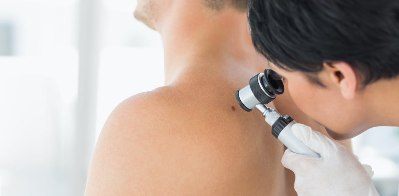
You have to to have common pores and skin examinations to catch any new melanomas early.
Writer equipped
If the node has no melanoma cells, you gained’t want any additional therapy apart from a six-monthly or yearly follow-up bodily examination. This checks for sudden recurrences by checking for swelling at your lymph nodes and any regarding signs reminiscent of unexplained weight reduction, ache or fatigue. This verify may even search for new major melanomas on the pores and skin.
Learn extra:
Solar harm and most cancers: how UV radiation impacts our pores and skin
If there’s a constructive lymph node, you is perhaps invited to take part in scientific trials with immunotherapy medicine. There may even be additional investigations to verify whether or not the melanoma has unfold additional, and in that case, the place.
These scale up from ultrasound checks of the remainder of the lymph nodes, to PET/CT scans of the remainder of the physique, and even MRI scans if there are indicators of unfold to the mind. If there are metastases, you’ll work with an oncologist to debate surgical removing and what drug therapy you may want.
Having one melanoma means you’re vulnerable to having extra sooner or later, so the ultimate a part of your therapy program will likely be common pores and skin checks along with your physician. Your GP may do these themselves, or refer you to a dermatologist or one other GP who has specialised in pores and skin most cancers.
You must also study to verify your individual pores and skin between visits, so you possibly can deliver a brand new or altering lesion to your physician’s consideration early.

Katie Lee receives funding from the NHMRC and The College of Queensland.
H. Peter Soyer is a shareholder of MoleMap NZ Restricted and e-derm seek the advice of GmbH, and undertakes common teledermatological reporting for each corporations. He’s a Medical Advisor for Canfield Scientific Inc, MoleMap Australia Pty Ltd, Blaze Bioscience Inc, and a Medical Advisor for First Derm. He holds an NHMRC MRFF Subsequent Technology Medical Researchers Program Practitioner Fellowship (APP1137127) and several other different NHMRC and MRFF grants. He’s a Board Member of Melanoma and Pores and skin Most cancers Trials Restricted and the Queensland Pores and skin and Most cancers Basis. He’s employed by The College of Queensland and works as Visiting Medical Officer at Metro South HHS.
Erin McMeniman doesn’t work for, seek the advice of, personal shares in or obtain funding from any firm or organisation that will profit from this text, and has disclosed no related affiliations past their educational appointment.